
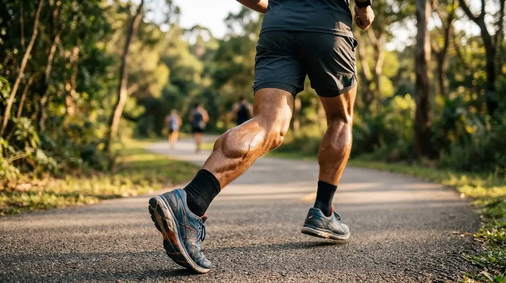
The calves are an essential part of the lower body, supporting everyday activities such as walking, running, climbing stairs, and maintaining balance. Located at the back of the lower legs, these muscles help control ankle movement and provide the force needed to push the body forward. Understanding calf anatomy and function can help you appreciate how your legs work during movement and exercise.
Although many people think of the calves as a single muscle, the region contains several muscles, tendons, nerves, and blood vessels that work together. The gastrocnemius and soleus are the two primary muscles responsible for much of the calf’s movement and strength. Their coordinated actions support stability, mobility, and efficient movement throughout daily activities.
Learning about the calves is useful for anyone interested in human anatomy, physical fitness, sports performance, or general muscle health. This guide explains the structure of the calf muscles, their functions, blood supply, nerve connections, and common conditions. It also explores how the calves contribute to walking, posture, circulation, and healthy lower-body movement.
1. What Are the Calves in Human Anatomy?
The calves are the muscular region located at the back of the lower legs, between the knees and ankles. They form the rounded posterior portion of the lower leg and contain muscles responsible for important ankle and foot movements. These muscles work closely with surrounding bones, tendons, ligaments, and nerves to support physical activities.
The calf region is primarily associated with the gastrocnemius and soleus muscles, which together form the triceps surae. These muscles attach to the heel through the Achilles tendon and produce much of the force required for plantar flexion. This movement occurs when the foot points downward or the heel lifts away from the ground.
Other muscles within the posterior lower leg assist with movement and stability. These include the plantaris, tibialis posterior, flexor hallucis longus, and flexor digitorum longus. Each has a different anatomical position and function, contributing to coordinated foot movements, balance, and the support needed during standing and walking.
2. Understanding the Main Calf Muscles
The calf muscle group contains several important structures, but the gastrocnemius and soleus perform most of the visible and functional work. Together, they generate force during walking, running, jumping, and standing on the toes. Their position behind the lower-leg bones allows them to influence ankle movement and support efficient body mechanics.
The gastrocnemius is the larger superficial muscle that forms much of the recognizable calf shape. It crosses both the knee and ankle joints, meaning it contributes to movements at both locations. The soleus lies underneath the gastrocnemius and plays a particularly important role in maintaining posture and controlling ankle position.
These two muscles work together but have different mechanical characteristics. The gastrocnemius contributes strongly to dynamic movements, while the soleus provides important endurance and postural support. Understanding these differences helps explain why calf muscles are involved in both powerful athletic activities and simple tasks such as standing comfortably.
3. Gastrocnemius Muscle Anatomy and Function
The gastrocnemius is a prominent calf muscle located near the surface of the posterior lower leg. It has two heads, commonly described as the medial and lateral heads, which originate from the lower portion of the femur. These muscle sections join and contribute to the Achilles tendon near the back of the ankle.
Because the gastrocnemius crosses the knee and ankle joints, it performs more than one movement function. Its primary role is helping produce plantar flexion at the ankle, allowing the heel to rise during walking and jumping. It also assists with knee flexion, although other muscle groups provide much of the force for bending the knee.
The gastrocnemius becomes especially active during movements that require powerful ankle extension and rapid changes in direction. Sprinting, jumping, and climbing stairs all involve coordinated calf muscle activity. Its contribution varies according to knee position, movement speed, and the amount of force required during a particular physical activity.
4. Soleus Muscle Anatomy and Function
The soleus is a broad, relatively flat muscle located beneath the gastrocnemius in the posterior lower leg. It originates mainly from portions of the tibia and fibula and contributes to the common tendon attaching to the heel. Although less visible than the gastrocnemius, it plays an essential role in lower-leg function.
Unlike the gastrocnemius, the soleus crosses only the ankle joint. Its primary function is plantar flexion, helping push the foot downward and supporting the body during standing. The muscle contains a relatively high proportion of fatigue-resistant fibers, making it particularly suited to sustained activity and postural control.
The soleus works continuously during many everyday movements, often without conscious attention. When standing, it helps control the body’s tendency to move forward at the ankle. During walking, it contributes to supporting body weight and controlling the movement of the lower leg over the planted foot.
5. Plantaris and Other Supporting Calf Muscles
The plantaris is a small muscle located in the posterior lower leg near the gastrocnemius and soleus. It typically has a short muscle belly and a long, thin tendon extending toward the heel. However, this muscle is naturally absent in some individuals and is not essential for normal lower-leg movement.
The plantaris provides a relatively minor contribution to ankle plantar flexion and knee flexion. Its mechanical role is much smaller than that of the gastrocnemius and soleus. Because of its long tendon and anatomical variability, the plantaris is also of interest in clinical anatomy and certain orthopedic procedures.
Deeper posterior leg muscles include the tibialis posterior and the long flexors of the toes. These structures support foot positioning, toe movements, and the stability of the foot during walking. Although they are not usually considered the main visible calf muscles, they are important components of the region’s overall functional anatomy.
6. How the Achilles Tendon Connects the Calf Muscles
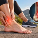
The Achilles tendon is a strong connective tissue structure located at the back of the ankle. It primarily connects the gastrocnemius and soleus muscles to the calcaneus, commonly known as the heel bone. This connection allows muscular force generated in the calf to influence movement of the foot and ankle.
When the calf muscles contract, tension travels through the Achilles tendon to the heel. This mechanical action helps produce plantar flexion, an essential movement during walking, running, and jumping. The tendon also stores and releases elastic energy during certain activities, improving movement efficiency and supporting repeated steps.
Despite its strength, the Achilles tendon can experience irritation or injury when exposed to loads beyond its current capacity. Gradual changes in activity and suitable recovery are important considerations for maintaining tendon health. Persistent pain, swelling, or sudden difficulty using the ankle should be assessed by a qualified healthcare professional.
7. What Are the Main Functions of the Calf Muscles?
The primary function of the calf muscles is plantar flexion, which involves pointing the foot downward at the ankle. This movement allows the heel to lift during walking and helps propel the body forward. It is also essential for activities such as running, jumping, cycling, and rising onto the toes.
Calf muscles also contribute to balance and postural stability. The soleus is especially important during standing because it helps control subtle movements at the ankle. The gastrocnemius provides additional force during activities requiring quick movement, changes in direction, or more demanding lower-body coordination.
Another important function involves controlling how the ankle moves while the foot remains in contact with the ground. During walking, the calf muscles help regulate the forward movement of the lower leg. This controlled action contributes to stable steps, efficient weight transfer, and coordinated movement between the foot, ankle, knee, and hip.
8. How the Calves Work During Walking and Running
Walking involves a coordinated sequence of movements between the hips, knees, ankles, and feet. The calf muscles become particularly important when the body moves over the supporting foot. They help control ankle motion and contribute to the push-off phase, allowing the opposite leg to move forward.
During running, the calves experience greater mechanical demands because movement involves faster loading and stronger forces. The gastrocnemius and soleus work with the Achilles tendon to support propulsion and manage contact with the ground. Their activity changes depending on running speed, terrain, footwear, and individual movement patterns.
Calf function also influences walking efficiency and overall lower-body coordination. When these muscles become fatigued or painful, people may change their walking patterns to compensate. Maintaining comfortable ankle mobility and adequate muscular capacity supports normal movement, although persistent gait problems may require professional assessment.
9. The Role of Calf Muscles in Balance and Posture
Balance depends on coordinated communication between the nervous system, muscles, joints, and sensory structures. The calf muscles contribute by producing small adjustments at the ankle when the body shifts forward or backward. These adjustments help maintain the body’s position over its supporting base during standing and movement.
The soleus plays an especially important role in upright posture because it helps resist forward movement of the lower leg. Its fatigue-resistant muscle fibers support sustained activity without requiring repeated powerful contractions. The gastrocnemius also contributes to stability, particularly when balance adjustments involve greater movement or force.
Good balance requires more than calf strength alone. Foot muscles, hip stabilizers, vision, and the vestibular system all contribute to maintaining a steady position. When balance difficulties occur regularly, evaluating the broader movement system is important rather than assuming that weak calves are the only possible explanation.
10. Calf Muscle Blood Supply and Nerve Connections
The calf muscles depend on a reliable blood supply to receive oxygen and nutrients during movement and rest. Important arteries serving the posterior lower leg include branches of the popliteal artery, particularly the posterior tibial and fibular arteries. These vessels support muscle function and help maintain healthy lower-leg tissues.
The tibial nerve provides the primary nerve supply to the gastrocnemius, soleus, plantaris, and several deeper posterior leg muscles. It carries motor signals that allow these muscles to contract during movement. The nerve also contributes to sensory functions through its branches, supporting communication between the lower leg and nervous system.
Problems affecting circulation or nerve function can influence calf strength, sensation, and movement. Symptoms may include weakness, numbness, unusual sensations, or discomfort during activity. Because several conditions can cause similar symptoms, ongoing neurological or circulatory concerns should be evaluated by a healthcare professional rather than diagnosed through symptoms alone.
11. How Calf Muscles Support Blood Circulation
The calf muscles also contribute to venous circulation through a mechanism commonly called the calf muscle pump. When these muscles contract, they compress nearby veins and help move blood upward toward the heart. One-way valves within healthy veins reduce backward blood flow and support this process.
Walking and ordinary ankle movements activate the calf muscle pump repeatedly throughout the day. This mechanism is especially important because blood returning from the lower legs must move against gravity when a person is upright. The interaction between muscle contractions and venous valves helps maintain efficient circulation in the lower extremities.
Prolonged immobility can reduce the frequency of calf muscle contractions and contribute to blood pooling in the legs. Regular comfortable movement may help support normal circulation during periods of sitting. However, unexplained swelling, persistent pain, or changes in skin color require medical attention because circulation problems can have several causes.
12. Common Calf Muscle Injuries and Conditions
Calf discomfort can develop from muscle strains, excessive loading, cramps, or irritation of nearby tendons. A calf strain occurs when muscle fibers are overstretched or injured during physical activity. These injuries can affect the gastrocnemius or soleus and may cause pain, tenderness, swelling, or difficulty performing normal movements.
Muscle cramps are another common source of calf discomfort, often involving sudden involuntary muscle contractions. They may occur during exercise, after prolonged activity, or while resting. Several factors can contribute, including muscle fatigue, certain medications, and underlying health conditions, although the exact cause is not always identifiable.
Not every painful calf is caused by a muscle problem. Nerve conditions, circulation disorders, and deep vein thrombosis can also produce lower-leg symptoms. Sudden unexplained swelling, warmth, or pain in one leg needs prompt medical assessment, while calf symptoms accompanied by chest pain or breathing difficulty require emergency care.
13. Understanding Calf Tightness and Limited Mobility
Calf tightness is commonly described as a sensation of stiffness or restricted movement at the back of the lower leg. It may develop after physical activity, prolonged inactivity, or repeated movement demands. Sometimes the sensation reflects temporary muscle fatigue, while other situations involve limitations in ankle mobility or underlying tissue irritation.
Restricted ankle dorsiflexion occurs when the foot has difficulty moving upward toward the shin. Calf muscle flexibility, joint structure, and other soft tissues can influence this movement. Limited dorsiflexion may affect squatting, stair navigation, or walking mechanics, although individual differences in joint mobility are normal.
Gentle movement and appropriate mobility exercises may help address ordinary stiffness when no injury is present. However, stretching should not be forced through pain or used as a substitute for medical evaluation. Persistent tightness, recurring discomfort, or noticeable movement restrictions may benefit from assessment by a qualified physical therapist or healthcare professional.
14. Ways to Maintain Healthy Calf Muscles
Maintaining healthy calves involves regular physical activity, comfortable joint mobility, and appropriate recovery. Walking, cycling, and other suitable activities naturally involve the lower-leg muscles. The goal is to support everyday movement capacity and muscular function rather than focusing on a particular appearance or achieving unrealistic physical standards.
Calf-strengthening exercises can help improve muscular capacity when performed with appropriate technique and gradual progression. Simple movements such as controlled heel raises are commonly used in general fitness and rehabilitation settings. Exercise selection and intensity should reflect individual ability, existing medical conditions, and any symptoms affecting the ankle or lower leg.
Recovery is equally important because muscles and tendons respond to repeated physical demands over time. Adequate sleep, balanced nutrition, hydration, and suitable rest between demanding activities support normal tissue function. People recovering from calf injuries should follow individualized healthcare guidance rather than relying on general exercise recommendations alone.
15. Why Understanding Calf Anatomy Matters
Understanding calf anatomy helps explain why the lower legs are involved in so many everyday movements. The gastrocnemius, soleus, Achilles tendon, and deeper muscles work together to support walking, standing, and coordinated ankle motion. Their combined activity contributes to mobility, stability, and the efficient transfer of force through the lower body.
Basic anatomical knowledge can also improve awareness of common lower-leg symptoms. Recognizing the difference between muscle function, tendon involvement, and possible circulation concerns encourages more informed decisions about physical activity and healthcare. However, knowing the location of a structure does not establish the cause of pain or replace professional diagnosis.
Calf anatomy is relevant to students, fitness enthusiasts, healthcare learners, and anyone interested in how the human body works. Understanding the relationship between muscles, nerves, tendons, and bones provides a clearer picture of lower-limb function. This knowledge supports informed conversations about movement, exercise, recovery, and long-term musculoskeletal health.
Conclusion
The calves are an important muscle region responsible for supporting movement, balance, posture, and ankle stability. The gastrocnemius and soleus form the main calf muscle group and work together through the Achilles tendon to produce plantar flexion. Additional muscles, nerves, blood vessels, and connective tissues contribute to coordinated lower-leg function.
Understanding calf anatomy helps explain how these structures support everyday activities such as walking, running, climbing stairs, and standing. The calves also play a role in venous circulation through the calf muscle pump, making regular movement valuable for normal lower-leg function. Proper activity, gradual strengthening, and adequate recovery help maintain healthy muscles and tendons.
Although occasional calf stiffness or discomfort may occur, persistent symptoms should not be ignored. Recognizing unusual pain, swelling, weakness, or mobility problems can help people seek appropriate medical advice. By understanding the structure and functions of the calves, you can develop a better appreciation of their essential role in healthy movement and overall lower-body function.
Frequently Asked Questions
What are the main muscles in the calves?
The gastrocnemius and soleus are the primary calf muscles, together forming the triceps surae. Other posterior lower-leg muscles, including the plantaris and tibialis posterior, contribute to movement and stability.
What is the main function of the calf muscles?
The main function of the calf muscles is ankle plantar flexion, which helps lift the heel and push the body forward. They also support walking, running, jumping, balance, and upright posture.
What is the difference between the gastrocnemius and soleus?
The gastrocnemius crosses the knee and ankle joints, contributing to plantar flexion and knee flexion. The soleus crosses only the ankle and plays an important role in sustained postural control.
Why do calf muscles become tight or painful?
Calf tightness or pain may result from muscle fatigue, strains, limited ankle mobility, or other health conditions. Persistent pain, unexplained swelling, or unusual symptoms should be assessed by a healthcare professional.
How can you maintain healthy calf muscles?
Regular walking, appropriate strengthening exercises, comfortable ankle mobility, and sufficient recovery can support healthy calf function. Activities should match individual abilities, and painful movements should not be forced.